How to identify corals with hitchhikers in a reef aquarium

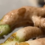
Carefully inspecting corals should be part of the regular husbandry routine for a reef aquarium so that any signs of soft tissue damage or discoloration can be identified and investigated as soon as possible.


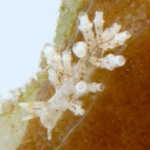
Early detection of the presence of hitchhikers is the most effective way of preventing an uncontrolled outbreak that can potentially harm the entire reef.


In the case of a mild infestation, the corals can suffer from chronic stress and will show signs of polyp retraction, pale patches on the soft tissue and increased mucus secretion. If not treated, it will become a severe infestation that can cause severe damage to corals, such as:

  • Direct damage to the soft tissue as a result of predation and feeding or by the extraction of proteolytic enzymes and chemo-toxins, eventually exposing the coral skeleton.
  • Reduced resistance against sedimentation, dehydration, UV-radiation, and bacterial infections as the hitchhikers feed on the corals mucus layer that protects it.
  • Nutrition deficiencies as the erosion of the mucus layer reduces the corals ability to capture dissolved and suspended organic materials.
  • Significant reduction in the amount of energy the coral receives from the photosynthetic zooxanthellae due to hundreds of creatures moving across the soft tissue and polyps, blocking the light.

Hitchhikers Identification Guide

Acropora-Eating Flatworms AEFW​

Red Acoel Flatworms

Where to look

All over the aquarium


What they look like

Tan, brown, red, coloured, flat worm, often with a red dot. Oval and slightly elongated with two tail-like appendages at their posterior, usually 3-6 mm in length.


What to look for

Flat worms covering every surface of the aquarium. In heavy infestation can obscure corals.

red-bug
red-bug

Red/Black Acro Bugs

Where to look

Acropora species


What they look like

0.5 – 1 mm yellow or golden copepod with a prominent red spot.


What to look for

Patches of tissue necrosis.

Parasitic copepod
Parasitic copepod

LPS parasitic copepod

Where to look

Acanthastrea, Micromussa, Favia & Favites


What they look like

0.5 – 1 mm yellow or white elongated copepod with a several lateral red spot.


What to look for

Copepods entrench between the coral polyps and produce mucus-sand cement that suffocate the polyp and inflict death.

Montipora-eating nudibranchs pale

Montipora-eating nudibranchs pale

Where to look

Montipora & Anacropora


What they look like

White to tan in color, up to 0.5 cm in length branch-like appendages along their back.


What to look for

Nudibranchs clusters line up along the edge of necrotic and bare skeleton.

Acropora-Eating Flatworms AEFW

Where to look

Acropora species


What they look like

Tan to opaque flatworm, mostly oval, 1-5 mm in length. Very difficult to detect on coral.


What to look for

Light infestation – small white patches of tissue necrosis (feeding scars) of approximately ~1 mm

Severe infestation – Severe damage to the soft tissue with massive skeleton exposure. eggs which are golden brown masses on the base or underside of white, dead coral tissue.

Montipora -Eating Flatworms

Where to look

Montipora species


What they look like

Tan to opaque flatworm, mostly oval, 1-5 mm in length. Very difficult to detect on coral.


What to look for

Light infestation – small white patches of tissue necrosis (feeding scars) of approximately ~1 mm

Severe infestation – Severe damage to the soft tissue with massive skeleton exposure. eggs which are golden brown masses on the base or underside of white, dead coral tissue

Watch the DipX coral dip video and learn how to safely introduce new corals into your tank: